
伊拉克硕大利什曼原虫银纳米颗粒的绿色合成与治疗

利什曼病是一组由利什曼原虫属(Leishmania)原生动物引起的传染性和非传染性严重寄生虫病。传播通过感染的雌性白蛉属(Phlebotomus)白蛉吸血而发生,白蛉被视为这些寄生虫的中间宿主。寄生虫的鞭毛前鞭毛体形态存在于白蛉体内,一旦被宿主巨噬细胞吞噬,就会转变为无鞭毛的无鞭毛体形态(1,2)。
利用微生物(3,4)、酶(5)、真菌(6)以及植物或植物提取物(7,8)合成纳米颗粒(NPs)的生物学方法,已被认为是化学和物理方法的可能的生态友好型替代方案。纳米颗粒绿色合成工艺的开发,正在发展成为纳米技术的一个重要分支,尤其是具有许多应用的银纳米颗粒(9-11)。化学合成方法会导致表面吸附一些有毒化学物质,这可能对医学应用产生不良影响。绿色合成比化学和物理方法更有优势,因为它具有成本效益、环境友好、易于放大以进行大规模合成,而且这种方法无需使用高压、能量、高温和有毒化学品(12,13)。银纳米颗粒因其诱人的物理化学性质,在生物学和医学领域发挥着重要作用。银制品长期以来被认为具有很强的抑制和杀菌作用以及广谱抗菌活性,几个世纪以来一直被用来预防和治疗各种疾病,最显著的是感染(14)。
NPs的粒径在抗菌效率中起着重要作用(15),因此最小粒径NPs的最强效果比最大粒径的更明显(16)。银纳米颗粒、金、壳聚糖和氧化金属的存在,会抑制贾第虫、利什曼原虫、疟原虫、弓形虫等一些寄生虫和昆虫幼虫的生长(17)。
材料与方法: AgNPs-TA的制备 在25 °C下的锥形瓶中,将5 mL鞣酸(TA)加入5 mL AgNO 3中。 混合物在强力磁力搅拌下保持5小时,直至出现棕色的AgNPs-TA胶体分散液,并避光保存直至使用。
纳米颗粒的表征 紫外-可见光谱在吸光度模式(200-900 nm范围)下使用双光束测定,以检查纳米颗粒的形成。粒径、形状和形态通过扫描电子显微镜(SEM)图像评估。
前鞭毛体的制备 从人体皮肤病变中分离出的硕大利什曼原虫(L. major)无鞭毛体,在含琼脂(4 mg/100 ml)和脱纤维兔血(10%)的Novy‐MacNeal‐Nicolle(NNN)培养基上培养。无鞭毛体转变为前鞭毛体样形态后,前鞭毛体在含100 μg/ml青霉素、100 μg/ml链霉素并添加10%胎牛血清(FCS)(Gibco Co.)的RPMI 1640培养基中继续生长,在28 °C培养箱中培养两周。在多次传代期间,寄生虫悬液在冷冻离心机中以4500 rpm离心沉淀10分钟,并去除上清液。这样,寄生虫在12天后进入固定生长期(稳定期),并准备好用于CL注射和诱导(18, 19)。
实验分组 感染硕大利什曼原虫的小鼠被随机分为三组,每组10只,具体如下:
第1组不作治疗,为对照组。 第2组在麻醉下接受剂量为2 mg/kg的AgNPs(20)。 第3组在麻醉下接受剂量为2 mg/kg的AgNPs-TA。
上述所有治疗均在第0、4、8和12天给予病灶部位。
评估疗效的参数 根据病灶和脾脏寄生虫负荷的变化记录治疗效果。每周两次测量病灶大小。首次治疗后处死小鼠,取其脾脏以确定活寄生虫的数量(21)。将动物的脾脏置于含完全RPMI‐1640培养基的6孔板中。脾组织匀浆后,将所有孔的内容物用完全RPMI‐1640培养基补足至200微升。所有样本分为三个重复。将制备好的板置于28 °C培养箱中10天。最后,根据显微镜观察,以系列稀释的方式报告阳性和阴性结果。
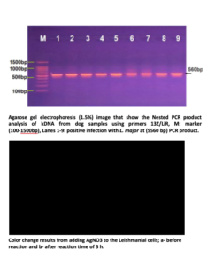
结果与讨论: 可靠且环保的金属纳米颗粒合成工艺的开发,是纳米技术应用领域的重要一步。使用化学合成方法时,一些有毒化学物质会吸附在表面,这可能对医学应用产生副作用。绿色合成比化学和物理方法更受青睐,因为它具有成本效益、环境友好、易于放大以进行大规模合成,而且这种方法无需使用高压、能量、高温和有毒化学品。因此,利用植物提取物的生物学方法而不是其他有毒方法来生产银纳米颗粒,是最好也最有效的(22,23)。一些金属纳米颗粒在可见光区域出现吸收带,这是由于电子的集体振荡与入射电磁辐射发生共振,称为表面等离子体共振(SPR)带(24)。对疑似皮肤利什曼病患者的六十份皮肤样本,通过显微镜观察检测利什曼原虫无鞭毛体,其中45份(75%)呈阳性;不过,NNN培养使42份样本(70%)长出了前鞭毛体。此外,结果显示,通过PCR方法,54份(90%)样本呈阳性;其中60%为硕大利什曼原虫(L.major),30%为热带利什曼原虫(L.tropica)。
硕大利什曼原虫细胞暴露于银离子时,显示出以425 nm为中心的明显且相当宽的紫外-可见吸收带,因为这种纳米颗粒分散良好,没有聚集。该吸收带的出现被归属为表面等离子体,对于粒径在2至100 nm的各种金属纳米颗粒,这一点已有充分记录(25)。SEM显示形成了平均粒径为35至40 nm的银纳米颗粒,颗粒之间有间距。紫外-可见吸收光谱已被证明对银胶体的形成相当敏感,因为银纳米颗粒由于表面等离子体激发而表现出强烈的吸收峰(26,27)。
寄生虫在AgNPs-TA存在下的出现,提示了一些可能的作用机制。以往的文献报道,银会攻击细菌的细胞膜表面,改变通透性、渗透平衡,从而改变细胞的代谢途径。银纳米颗粒还可能与DNA结合,阻止复制(28-32)。TA包覆的金和银纳米颗粒的SPR带,与文献报道值(分别为520和380-450 nm)相比发生了红移(33,34)。这种红移效应很可能是由纳米颗粒与表面保护性TA多酚之间的超分子相互作用引起的,它改变了电子转移能量。除形状、尺寸和材料类型外,SPR带的位置高度依赖于周围介质的介电常数,因为它会改变电子在NMNPs表面的共振频率(35-37)。
结论: 用鞣酸合成的银纳米颗粒具有作为抗利什曼原虫治疗手段的潜在应用。此外,使用天然化合物有助于开发清洁、无毒、生物相容且对环境无害的方法来合成贵金属纳米颗粒。
参考文献:
-
Carneiro SMP, Carvalho FAA, Santana LCLR, Sousa APL, Moita-Neto JM and Chaves MH. The cytotoxic and antileishmanial activity of extracts and fractions of leaves and fruits of Azadirachta indica. Biol Res., 2012; 45: 111-116.
-
Kevric I, Cappel MA and Keeling JH. New World and Old World Leishmania Infections: A Practical Review. Dermatol Clin., 2015; 33: 579-593.
-
Klaus T, Joerger R, Olsson E, Granqvist CG. Silver-based crystalline nanoparticles, microbially fabricated. J. Proc. Natl. Acad. Sci.USA., 1999; 96: 13611-13614.
-
Konishi Y, Uruga T. Bioreductive deposition of platinum nanoparticles on the bacterium Shewanella algae. J Biotechnol ., 2007; 128:648-653.
-
Willner I, Baron R, Willner B. Growing metal nanoparticles by enzymes. J. Adv. Mater., 2006; 18:1109-1120.
-
Vigneshwaran N, Ashtaputre NM, Varadarajan PV, Nachane RP, Paraliker KM, Balasubramanya RH. Biological synthesis of silver nanoparticles using the fungus Aspergillus flavus. Mater Lett., 2007; 61:1413-1418.
-
Shankar SS, Ahmed A, Akkamwar B, Sastry M, Rai A, Singh A. Biological synthesis of triangular gold nanoprism. Nature. 2004; 3:482.
-
Ahmad N, Sharma S, Singh VN, Shamsi SF, Fatma A, Mehta BR. Biosynthesis of silver nanoparticles from Desmodium triflorum: a novel approach towards weed utilization. Biotechnol. Res. Int., 2011; 454090 (1-8).
-
Armendariz V, Gardea-Torresdey JL, Jose Yacaman M, Gonzalez J, Herrera I,Parsons JG. Proceedings of Conference on Application of Waste Remediation Technologies to Agricultural Contamination of Water Resources. Kansas City Mo, USA. 2002.
-
Kim BY, Rutka JT, Chan WC. Nanomedicine. N. Engl. J. Med., 2010; 363 (25): 2434-2443.
-
Kyriacou SV, Brownlow WJ, Xu XN. Using nanoparticle opti chitosan assay for direct observation of the function of antimicrobial agents in single live bacterial cells. Biochemistry. 2004; 43:140-147.
-
Singh A, Jain D, Upadhyay MK, Khandelwal N, Verma HN. Green synthesis of silver nanoparticles using Argimone mexicana leaf extract and evaluation of their antimicrobial activities. Digest J. Nanometer. Biostruct., 2010; 5 (2): 483-489.
-
Jain D, Kumar Daima H, Kachhwaha S, Kothari SL. Synthesis of plant-mediated silver nanoparticles using Papaya fruit extract and evaluation of their antimicrobial activities. Digest J. Nanomater. Biostruct., 2009; 4(3):557-563.
-
Shankar, S.S., Rai, A., Ankamwar, B., Singh, A., Ahmad, A., Sastry, M.. Nat. Mater., 2004; 3, 482–488.
-
Adams, LK, Lyon, DY, Alvarez, PJ. Comparative eco-toxicity of nanoscale TiO2, SiO2, and ZnO water suspensions. Water Res., 2006; 40: 3527-32.
-
Lu, Z, Rong, K, Li, J, Yang, H, Chen, R. Size-dependent antibacterial activities of silver nanoparticles against oral anaerobic pathogenic bacteria. J. Mater. Sci. Mater. Med., 2013; 24:1465-71.
-
Elmi, T, Gholami, S, Fakhar, M, Azizi, F. A Review on the most of nanoparticles in the treatment of parasitic infections. J. Mazand. Univ. Med. Sci. 2013; 23, 102:126-33.
-
Sazgarnia A, Zabolinejad N, Layegh P, Rajabi O, Berenji F, Javidi Z, et al. Antileishmanial activity of liposomal clarithromycin against Leishmania major Promastigotes. Iran J Basic Med Sci 2012; 15:1210‐1214.
-
Mohebali M, Rezayat MM, Gilani K, Sarkar S, Akhoundi B, Esmaeili J, et al. Nanosilver in the treatment of localized cutaneous leishmaniasis caused by Leishmania major (MRHO/IR/75/ER): an in vitro and in vivo study. DARU 2009;4:285‐289.
-
Xue Y, Zhang S, Huang Y, Zhang T, Liu X, Hu Y, et al. Acute toxic effects and gender‐related biokinetics of silver nanoparticles following an intravenous injection in mice. J Appl Toxicol 2012; 32:890‐899.
-
Buffet P, Sulahian A, Garin Y, Nassar N, Derouin F. Culture microtitration: a sensitive method for quantifying Leishmania infantum in tissues of infected mice. Antimicrob Agents Chemother 1995; 39:2167‐2168.
-
Jain D, Kumar Daima H, Kachhwaha S, Kothari SL. Synthesis of plant-mediated silver nanoparticles using Papaya fruit extract and evaluation of their antimicrobial activities. Digest J. Nanomater. Biostruct., 2009; 4(3):557-563.
-
Vyom P, Rashmi P, Bechan S, Avinash CP. Parthenium leaf extract mediated synthesis of silver nanoparticles: a novel approach towards weed utilization. Digest J. nanometer. Biostructures., 2009; 4(1):45-50.
-
Melo JR MA, Santos LSS, Goncalves MC and Nogueira AF. Preparação de nanopartículas de prata e ouro: um método simples para a introdução da nanociência em laboratório de ensino. Quim Nova., 2012; 35: 1872-1878.
-
El-Raheem A, El-Shanshoury AR, ElSilk SE, Ebeid ME. Extracellular biosynthesis of silver nanoparticles using Escherichia coli ATCC 8739, Bacillus subtilis ATCC 6633, and Streptococcus thermophilus ESh1 and their antimicrobial activities. ISRN Nanotechnol.,12011; -7.
-
Gao, J., K. Powers, et al. "Influence of Suwannee River humic acid on particle properties and toxicity of silver nanoparticles." Chemosphere., 2012; 89 (1): 96-101.
-
Abdulsadah A. Rahi1, Magda A. Ali and Alaa H. Al-Charrakh. Biosynthesis of silver nanoparticles by Leishmania tropica. Afr. J. Biotechnol., 2013; 12 (48): 6718-6722.
-
Jebali A and Kazemi B. Nano-based antileishmanial agents: A toxicological study on nanoparticles for future treatment of cutaneous leishmaniasis. Toxicol In Vitro. 2013; 27: 1896-1904.
-
Chen M, Yang Z, Wu H, Pan X, Xie X and Wu C. 2011. Antimicrobial activity and the mechanism of silver nanoparticle thermosensitive gel. Int J Nanomed 6: 2873-2877.
-
Perni S, Piccirillo C, Pratten J, Prokopovich P, Chrzanowski W, Parkin IP and Wilson M. The antimicrobial properties of light activated polymers containing methylene blue and gold nanoparticles. Biomaterials. 2009; 30: 89-93.
-
Allahverdiyev AM, Abamores, Bagirova M, Ustundag CB, Kaya C Kaya F and Rafailovich M. Antileishmanial effect of silver nanoparticles and their enhanced antiparasitic activity under ultraviolet light. Int J Nanomed., 2011; 6: 2705-2714.
-
Duran N, Marcato PD, Contird, Alves OL,Costa FTM and Brocchi M. Potential use of silver nanoparticles on pathogenic bacteria, their toxicity and possible mechanisms of action. J Braz Chem Soc., 2010; 21: 949-959.
-
Sharifi N, Dabirian A, Danaei D and Taghavinia N. Aggregates of plasmonic nanoparticles for broadband light trapping in dye-sensitized solar cells. J Opt-UK., 2015;18: 1-7.
-
Silva ATB, Coelho AG, Lopes LCS, Martins MVA, Crespilho FN, Merkoci A and Silva WC. Nano-assembled supramolecular films from chitosan-stabilized gold nanoparticles and Cobalt (II) phthalocyanine. J Braz Chem Soc., 2013; 24: 1237-1245.
-
Song and Jing. Insight into the localized surface plasmon resonance property of core-satellite nanostructures: Theoretical prediction and experimental validation. J Colloid Interf Sci., 2017; 505: 373-382.
-
Martinsson, Otte MA, Shahjamali MM,Sepulveda B and Aili D. Substract effect on the Reffractive Index sensity of silver nanoparticles. J Phys Chem C., 2014; 118: 24680-24687.
-
Mahmoud MA, Chamanzar M, Adibi A and Elseyed MA. Effect of the dielectric constant of the surrounding medium and the substrate on the surface plasmon resonance spectrum and sensitivity factors of highly symmetric systems:silver nanocubes. J Am Chem Soc., 2012; 134: 6434-6442.
Abdulsada A. Rahi 1*、Magda A. Ali 2和Zaid A. Abdulabbas(伊拉克库特市瓦西特大学理学院生物系(Department of Biology, College of Science, Wasit University, Kut, Iraq.)) (伊拉克库特市瓦西特大学医学院(College of Medicine, Wasit University, Kut, Iraq))
由英文原文自动翻译. 阅读原文