
Mass General Brigham AI predicts cancer survival risk from a facial photo
Researchers design foundation models FAHR-FaceAge and FAHR-FaceSurvival to assess biological frailty and patient mortality from a single digital snapshot
By Giovanni Sapere
2 min readHuman-Edited, AI-Assisted
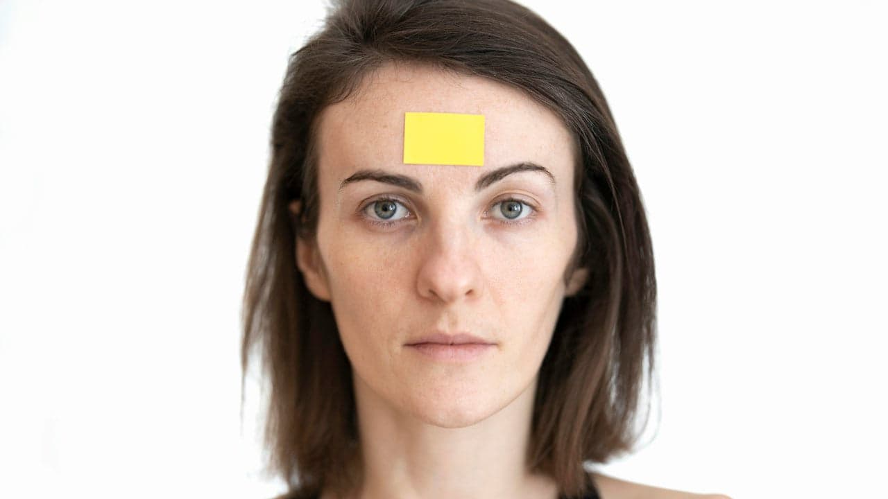
Scientists at the Mass General Brigham Cancer Institute have developed an artificial intelligence tool capable of predicting a patient's biological age and cancer treatment survival odds from a single facial photograph. The clinical breakthrough provides oncologists with a non-invasive method to evaluate physical reserve before ordering aggressive interventions such as radiotherapy, systemic therapy or surgery. Published in The Lancet Digital Health, the project shows how digital facial biomarkers can measure patient frailty without requiring exhausting clinical tests.
From facial pixels to clinical biomarkers
Determining how well a patient will endure cancer treatment traditionally depends on subjective clinical judgment and exhausting physical tests. To provide an objective metric, researchers created FAHR-Face, short for Foundation Artificial Intelligence for Health Recognition, as reported by New Atlas. The self-supervised machine learning model uses masked autoencoders trained on over 42 million photos of faces, teaching the system to reconstruct obscured features and capture structural information that correlates with general health.
“To our knowledge, this is the first demonstration that an AI model can directly predict cancer survival from a simple photograph.”
— Hugo Aerts, PhD, director of the AI in medicine program at Mass General Brigham
Two algorithms for age and survival
The researchers fine-tuned the core architecture into two complementary diagnostic instruments: FAHR-FaceAge and FAHR-FaceSurvival. According to data reported by New Atlas, FAHR-FaceAge calculates physiological aging rather than chronological years, achieving an average error margin of 5.1 years. Crucially, the model remained robust against confounding elements such as makeup, glasses, varying illumination and facelifts, showing only minor discrepancies in subjects who had undergone rhinoplasty. Patients appearing five or more years older than their chronological age faced a 21% higher risk of mortality, whereas those appearing five years younger exhibited a 17% higher survival rate.
Mapping mortality risk during cancer therapy
The survival tool was evaluated on facial images of more than 34,000 cancer patients undergoing radiotherapy, followed by validation cohorts of nearly 4,000 and 5,000 individuals. As detailed by New Atlas, FAHR-FaceSurvival successfully distinguished risk tiers: patients placed in the highest mortality category experienced more than triple the death rate of those in the lowest bracket. Within the highest-risk segment, 52% of patients died within 90 days, compared to only 2.1% in the lowest-risk group. Heatmaps revealed that FAHR-FaceSurvival analyzes the zone below the eyes and along the nasal bridge, whereas FAHR-FaceAge focuses primarily on the temples, nasolabial folds and the forehead above the eye sockets.
Clinical integration and public evaluation
By extracting physiological signals from consumer-grade photographs, this innovation could help oncologists tailor radiation dosages or choose less toxic alternatives for frail individuals. Co-senior author Hugo Aerts and radiation oncologist Raymond Mak noted that the foundation-model structure allows teams to develop specialized diagnostic markers without building each neural network from scratch. While researchers have opened an expanded online trial for public evaluation, clinical teams are preparing further assessments to determine how the automated visual biomarker can be safely integrated into clinical oncology workflows, as documented by New Atlas.
Read also on The Patent: Lab-Grown Neurons Decode Braille and Steer Robot Dogs