
Rockefeller University Engineers TrUDI Platform for Deep Brain Imaging
A redesigned three-photon microscope records neural activity in intact mouse brains at single-cell resolution.
By Letizia Giugliano
2 min readHuman-Edited, AI-Assisted
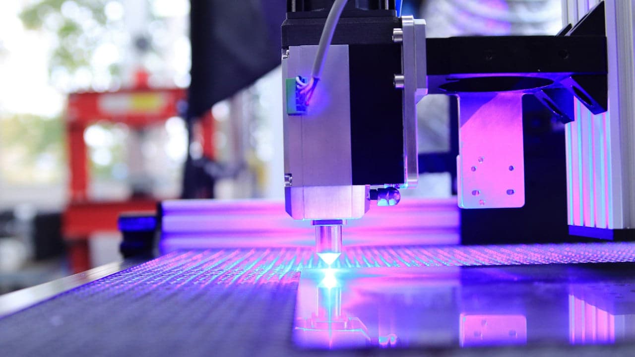
Biophysicists at Rockefeller University have developed a microscopy system that records neural activity deep within living brain tissue without requiring the surgical removal of overlying layers. The technical innovation, described in Nature Methods, boosts three-photon imaging efficiency by roughly 250 times while protecting delicate neural structures from heat damage.
Rebuilding three-photon optics from the ground up
Imaging single neurons inside living tissue poses a persistent physical challenge because mammalian brain structures scatter light. As optical beams travel deeper into tissue, scattered light creates background glow that masks cellular activity. Standard two-photon microscopes usually reach depths of roughly 0.8 millimeters, while conventional three-photon setups had reached functional recording limits of about 1.2 millimeters, falling short of the theoretical 2.3-millimeter physical threshold in mouse tissue, according to reporting by MedicalXpress.
To bridge that gap, a research team led by Alipasha Vaziri, head of the Laboratory of Neurotechnology and Biophysics at Rockefeller University, developed an optical invention named Three-photon Ultra-Deep Imaging, or TrUDI. Instead of adapting existing commercial components, the group modeled optical propagation limits computationally and built the hardware configuration from scratch.
“We wanted to take a clean-slate approach.”
— Alipasha Vaziri, Head of the Laboratory of Neurotechnology and Biophysics at Rockefeller University
Higher pulse energy and targeted cell scanning
Multiphoton excitation requires high laser intensity, but increasing average laser power overheats and harms living brain cells. The engineering team solved this operational barrier by delivering fewer laser pulses that carry substantially higher individual pulse energy, keeping the overall average thermal load within safe thresholds.
The researchers also modified how the microscope focuses light and scans across tissue. The system incorporates an excitation mechanism that detects individual cells and fires the laser beam only when sweeping over those target neurons. This selective scanning approach reduces total tissue exposure to laser radiation by up to 80 percent during extended imaging experiments, as detailed by MedicalXpress.
Capturing intact hippocampus and striatum signals
The custom prototype platform achieved a 250-fold increase in imaging efficiency compared with prior three-photon configurations. That performance enabled researchers to record functional activity across the entire mouse cortex and inspect deeper structures such as the hippocampus and the striatum without removing overlying cortical brain tissue.
Experimental trials documented in the study tracked neuronal activity in mice expressing fluorescent indicators including GCaMP8m and green fluorescent protein Thy1-EGFP. The team demonstrated volumetric imaging across a field of view measuring 230 by 230 by 1,680 cubic micrometers, capturing individual transient cellular signals that were previously obscured by optical scattering, according to MedicalXpress—marking a significant biomedical innovation for deep-tissue neuroimaging.
Read also on The Patent: IBM Links Two Quantum Chips with Superconducting Cables · From Prototype to Deployment: The Regulatory and Engineering Barriers in Health Innovation