
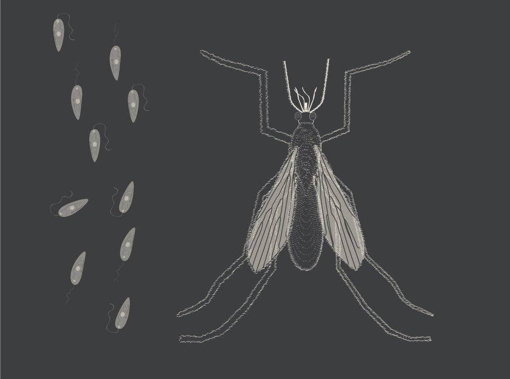
Leishmaniasis is a parasitic diseases induced by 20 dif-ferent species of Leishmania. Leishmaniasis is reported from 88 countries and estimated that 350 million world-wide are at risk of acquiring one form of the diseases, and 12 million are infected with annual incidence rate of about 1.5 to 2 million. According to WHO estimates, 90% of cutaneous cases occur in six countries. Leishmaniasis depends upon causative agent and host genetic back-ground presents various manifestations ranging from a self healing lesion to a lethal systemic form of the disease. Two most common clinical forms of the disease Cutaneous Leishmaniasis (CL) and visceral leishmaniasis (VL) are mainly seen in 14 of the 22 countries of Eastern Mediterranean Region (EMRO) (Postigo, 2010). Traditionally, Leishmania parasites are directly detected by microscopic examination of clinical specimens. However, in an endemic area, CL can generally be diagnosed by its clinical appearance alone (Kaur et al., 2003). Nanotechnology is the study of controlling matter on an atomic and molecular scale. Generally, it deals with structures of the size of 100 nm or smaller in at least one dimension, and involves developing materials or devices within that size (Kim et al., 2010). Biological methods of nanoparticles synthesis using microorganisms (Klaus et al., 1999; Konishi and Uruga, 2007), enzymes (Willner et al., 2006), fungus (Vigneshwaran et al., 2007), and plants or plant extracts (Shankar et al., 2004; Ahmad et al., 2011) have been suggested as possible eco-friendly alternatives to chemical and physical methods. The development of green processes for the synthesis of nanoparticles is evolving into an important branch of nanotechnology especially silver nanoparticles, which have many applications (Armendariz et al., 2002; Kim et al., 2010; Kyriacou et al., 2004). Chemical synthesis methods lead to presence of some toxic chemical absorbed on the surface that may have adverse effect in the medical applications.
Green synthesis provides advancement over chemical and physical method as it is cost effective, environment friendly, easily scaled up for large scale synthesis and in this method there is no need to use high pressure, energy, temperature and toxic chemicals (Singh et al., 2010; Jain et al., 2009).
In recent years, silver nanoparticles (Ag-NPs) have attracted considerable attention for medical and chemical applications due to their exceptional properties including antibacterial activity, high resistance to oxidation and high thermal conductivity (Feng et al., 2006; Lee, 2007; Soni and Prakash, 2011). Recently, findings have demon-strated that nanosilver has anti-inflammatory effects and increases wound healing and dressings of wounds. If these results are confirmed in vivo, nanosilver may be appropriate for ulcer treatment (Karla et al., 2010).
The aim of the present study was to evaluate the synthesis of silver nanoparticles using Leishmania tropica and determine the characteristics of produced material. This study is the first record of biosynthesis of silver nanoparticles by L. tropica in Iraq.
MATERIALS AND METHODS
Samples collection
Forty four (44) patients were selected for isolation of Leishmania species from their cultures. Skin scrapings from the lesion were obtained and smears prepared on a slide, stained with Giemsa and examined microscopically for presence of amastigotes. Bacterial contamination of Leishmania cultures was minimized by cleaning lesions with 70% methanol and local debridement before obtaining specimens. At least two Giemsa-stained slides for each patient were prepared for microscopic examination and cultured.
Culture
The samples were aspirated from the edge of the skin lesions and cultured in liquid phase (normal saline) of Novy MacNeal Nicolle (NNN) media. The culture was incubated at 25°C and checked for growth of Leishmania promastigotes for 28 days. Penicillin-G and streptomycin were added to the phosphate buffered saline (PBS) solution utilized in the NNN media culture (Eisenberger and Jaffe, 1999; Farahmand et al., 2008).
PCR
PCR assay was performed according to the manufacturer’s protocol (Sinagen, Iran) with the final volume of 25 μL of each PCR reaction. PCR amplification was carried out in a DNA Thermal Cycler (Master cycler gradient, San Leonardo, Canada) based on the following conditions: initial denaturation (95°C, 3 min; 63°C, 30 s; 72°C, 60 s) 1 cycle followed by 35 cycles including denaturation (93°C, 20 s), annealing (63°C, 20 s) and extension (72°C, 40 s). Finally, 10 μL of amplified samples without adding loading buffer were loaded in a 2% agarose gel containing 0.5 mg/ml ethidium bromide in electrophoresis and the products were visualized by ultraviolet (UV) transillumination.
Synthesis of Ag-NPs
The cell culture of L. tropica was kept under stirring (5,000 rpm) (5 g) for 5 min at 28°C till it were separated from the broth culture then the settled cells were washed with distilled water three times. One gram (1 g) of wet cell mass was then resuspended separately in 0.001 M AgNO3 solution at pH 5.4 to 6.0. The total mixture was left in room temperature (25°C) for 3 h, and the reaction carried out. For characterization, nanoparticle powders were diluted in pure acetone and the prepared suspension was ultrasonicated. The biotransformation was routinely monitored by visual observation of the biomass as well as measurement of the UV–Vis spectra from the leishmanial cells. The positive reaction appear as formation of a yellow- brown color of the medium due to the reduction of silver ions and production of silver nanoparticles in the medium. The scanning electron microscopy used SEM grids were pre-pared by taking small amount of sample powder on a copper coated grid and dried under lamp. The silver nanoparticles appeared as spherical in shape and the average size was from 35 to 40 nm with inter-particle distance.
RESULTS AND DISCUSSION
Diagnosis of L. tropica by PCR, culture and microscope
44 patients were detected for Leishmania amastigotes by microscopic observation out of which, 38 (86.4%) were positive; however, the NNN culture led to the growth of promastigotes in 40 samples (90.1%). Also, the results shows that all of the 10 samples were positive (100%) from the PCR assay .
UV–vis spectroscopy and SEM
- L. tropica cells when exposed to silver ions showed a distinct and fairly broad UV–Vis absorption band centered at 425 nm because this nanoparticle was well dispersed without aggregation. The appearance of this band, which was assigned to a surface plasmon, is well documented for various metal nanoparticles with sizes ranging from 2 to 100 nm (El-Raheem et al., 2011). SEM showed the formation of silver nanoparticles with an average size of 35 to 40 nm with inter-particle distance. The shapes of Ag-NPs proved to be spherical. These results confirm the presence of primary and secondary amines bonds; C=O, N=O, C=N and COOH bonds of proteins as cap-ping and stabilizing agent on the nanoparticles surface (Fatemeh and Bahram, 2012; Ramezani et al., 2012).
Development of reliable and eco-friendly process for the synthesis of metallic nanoparticles is an important step in the field of application of nanotechnology. Chemical synthesis methods lead to presence of some toxic chemical absorbed on the surface that may have adverse effect in the medical applications. Biosynthesis provides advancement over chemical and physical method as it is cost effective, environment friendly, easily scaled up for large scale synthesis and in this method there is no need to use high pressure, energy, temperature and toxic chemicals. Therefore, there is most need of silver nanoparticles synthesized by biological methods of plant extract instead of other toxic methods (Singh et al., 2010; Jain et al., 2009; Vyom et al., 2009). The present study confirmed that L. tropica is capable of producing silver nanoparticles within 3 h in contrast to other researchers who found that the main problem in the biological nano-particles is the slow rate of production (Narayanan and Sakthivel, 2010). This approach towards synthesis of silver nanoparticles has many advantages such as pro-cess scaling up, economic viability and safe way to produce nanoparticles. Parasitic diseases (like malaria, leishmaniasis, trypanosomiasis) are major health pro-blems around the globe (Edward and Krishna, 2004). Antiparasitic chemotherapy is the only choice of treat-ment of these parasitic infections. The reason for this is that these infections do not elicit pronounce immune response hence effective vaccination may not be possible (Watkins, 2003).
The main reason for using Ag-NPs in this study was their capacity to produce reactive oxygen species (ROS), which Leishmania parasites are known to be susceptible to. Recent studies showed that their wide surface areas, small sizes, and their ability to bind sulfur- and phosphorus-containing groups may lead to an increase in their antileishmanial effects. It was observed that Ag-NPs decreased the metabolic activity and proliferation values of parasites compared with the control groups (Allahverdiyev et al., 2011). This inorganic nanoparticle has a distinct advantage over conventional chemical anti-microbial agents. The most important problem caused by the chemical antimicrobial agents is multidrug resistance. Therefore, an alternative way to overcome the drug resis-tance of various microorganisms is needed desperately, especially in medical devices, etc. Ag ions have been used for decades as antimicrobial agent in various fields because of their growth-inhibitory capacity against micro-organisms (Kim et al., 2007). Our results are in agree-ment with other results in different parts of the world (Allahverdiyev et al., 2011; Singh et al., 2010; Kim et al., 2007), and disagrees with others (Narayanan et al., 2010; Sondi and Salopek-Sondi, 2004). The nanoparticles were primarily characterized by UV-vis spectroscopy, which was proved to be a very useful technique for the analysis of nanoparticles.
The reduction silver ions and formation of stable nanoparticles appeared quickly within 3 h of reaction making it one of the fastest bioreducing methods to produce silver nanoparticles (Kim et al., 2007). Reduction of silver ions present in the aqueous solution of silver complex during the reaction with the cell culture of L. tropica observed by the UV-Vis spectroscopy revealed the presence of silver nanoparticles may be correlated with the UV-Vis spectra. UV-Vis spectroscopy is well known to investigate the shape and size of nanoparticles. The scanning electron microscopy analysis confirmed the bioreduction of Ag+ ions to silver nanoparticles as well as morphological dimensions of Ag-NPs demonstrated that between 35 to 40 nm with inter-particle distance and they have irregular spherical shape. These findings are in agreement with those of other researchers (Fatemeh and Bahram, 2012; Ramezani et al., 2012; Ponarulselvam et al., 2012). This study concluded that the efficiency of L. tropica for synthesis of silver nanoparticles was found to be higher; also this method was cost effective and easily scaled up for large scale synthesis.
Written by Prof. Dr. Abdulsada A. Rahi and Dr. Magda A. Ali (Department of Biology, College of Science, Wasit University, Iraq)