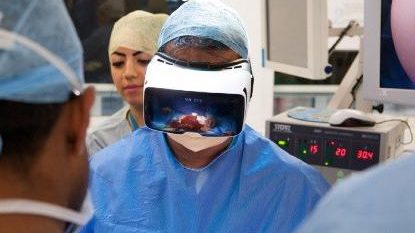
The project, which involves 12 European partners, develops a new surgical navigator that uses an innovative visor as a medical surgeon’s aid
The world’s first augmented reality surgery was a great success.
To announce it, the Policlinico Sant’Orsola in Bologna where the surgeon saw virtual elements in front of him during a maxillo-facial surgery.
Specifically, the operation consisted in resecting and repositioning a patient’s jaw and jaw in order to restore the functions of the bite: with the new technology it was possible to resect and reposition the jaw and jaw.
“This was made possible thanks to a state-of-the-art augmented reality viewer that the surgeon wore during the operation” says the University of Pisa, which coordinates the European project and created VOSTARS, the new generation viewer used by the Bolognese surgeon during the operation.
“Thanks to the use of the visor – explains Giovanni Badiali, project manager at the Policlinico Universitario Sant’Orsola in Bologna, who performed the operation –, before the operation, me and the whole team in the room, we visualized in augmented reality the anatomy of the facial skeleton, jaws and cutting lines. Subsequently, during the operation, the device made it possible to visualize a 3D dotted line directly on the patient’s bone, thus showing the path to follow with the surgical instrument. With the help of the viewer – he concludes – we were able to cut the jaw with the required precision“.
With the VOSTARS project (Video and Optical See Through Augmented Reality Surgical Systems) augmented reality enters the operating rooms.
The project, which involves 12 European partners, aims to develop a new surgical navigator using an innovative visor able to provide the surgeon with the vision of the operating room, specific information about the patient and more general information about the organs involved in the operation. All this is possible thanks to a video camera able to combine the images in front of the surgeon with the radiological images of the patient. Moreover, during the phases of the operation where accurate virtual guidance is not required, the viewer can become transparent allowing the surgeon to have a natural view of the operating field directly with his own eyes, as explained by Vincenzo Ferrari, biomedical engineer at the Department of Information Engineering of the University of Pisa and coordinator of the European team that designed the innovative viewer:
“To create VOSTARS we had to solve very complex problems, which mainly concern hand-eye coordination and coherence between the real and virtual image temporally, spatially and in terms of focus. It is obvious that if the surgeon has to follow a virtual cutting line these must appear in the right place and at the right time of surgery, but getting it is not trivial. Moreover, the virtual guide and the patient must be able to be focused at the same time to allow the surgeon to follow it with the scalpel – he concludes -. The viewers currently on the market make some digital content available directly in the field of vision, such as the three-dimensional image of the organ to be operated. Usually the physician displays these virtual images, obtained from X-ray scanners (such as CT and MRI scans), before the operation, to help him in the preparation of the operation. It has never happened until now, however, that a viewer was used to guide the actual surgical act“.