
Fluorescent Probes Reveal Fluid Barriers Inside Living Human Nuclear Pores
A study in Nature shows how sugar groups and transport receptors keep the cellular nanocanal flowing rather than hardening into a solid gel.
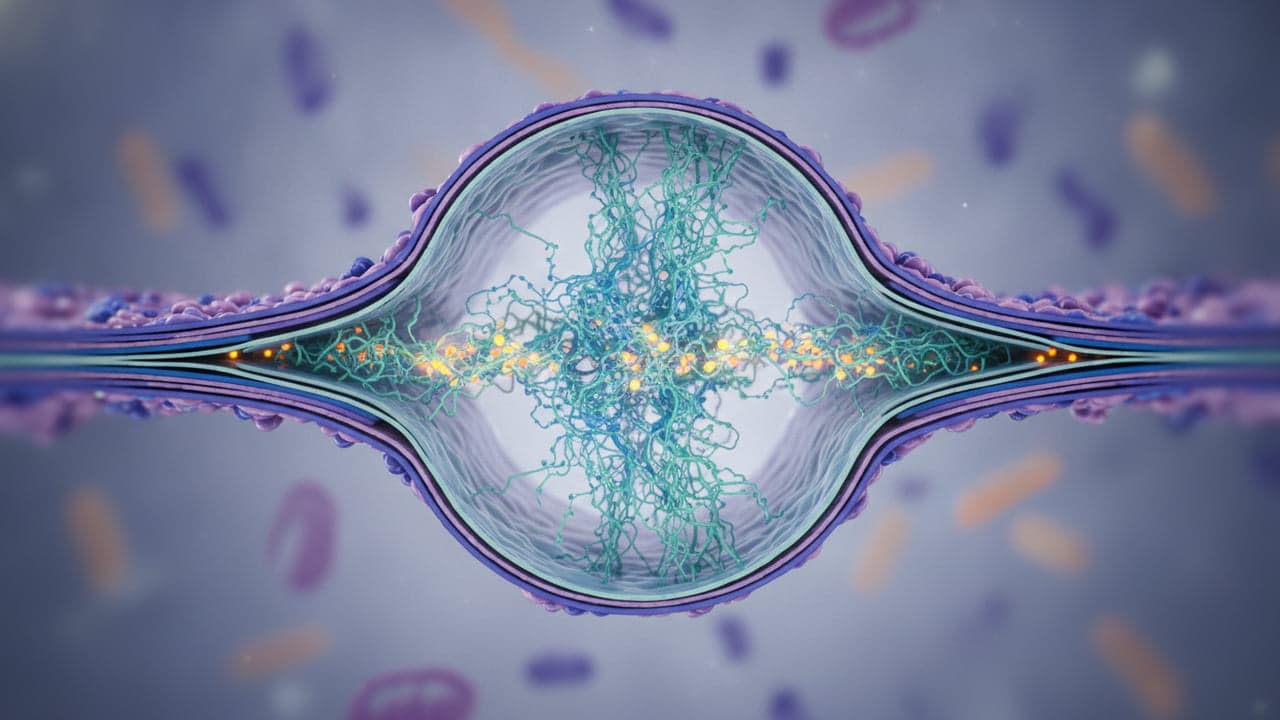
Researchers at Johannes Gutenberg University Mainz, the Max Planck Institute for Biophysics and Wuhan University have measured the protein dynamics inside human cell nuclear pores for the first time. The biophysical innovation shows that the central nanocanal remains in a fluid-like state rather than solidifying into a gel. The findings, published in Nature, solve a long-standing debate over how cells selectively filter tens of thousands of molecular visitors each minute.
Tracking protein motion in living nanocanals
Human cell nuclei protect their genetic information behind a double membrane perforated by thousands of microscopic channels. Transport through these gates is managed by an internal protein mesh, but scientists previously disagreed on whether that mesh behaved as a stretched polymer, a liquid condensate or a rigid gel. To test the structure in intact human cells through a new methodological innovation, a research team led by Johannes Gutenberg University Mainz (JGU), the Max Planck Institute for Biophysics (MPI-BP) in Frankfurt am Main and Wuhan University attached tiny glowing probes directly to pore proteins, as reported by Phys.org.
By tracking the mobility of these fluorescent dyes, the researchers could record the physical state of the canal directly. The dyes moved rapidly inside active cells, confirming that the internal channel operates like a liquid. Professor Edward Lemke, professor of synthetic biophysics at JGU who led the study alongside Professor Gerhard Hummer from the MPI-BP, explained that the high speed of the optical markers demonstrated a fluid-like environment inside the living pore.
“For the first time, we have been able to measure intrinsic protein dynamics within the nanocanal of a living cell and demonstrate that the interior of this canal is fluid-like”
— Professor Edward Lemke, Johannes Gutenberg University Mainz
Why cellular filters avoid solidifying
The dynamic behaviour observed in living cells contrasted sharply with tests conducted in test tubes. When isolated pore proteins were studied in vitro without cellular factors, the material consistently stiffened after roughly two hours into a dense structure resembling a gummy bear or the protein aggregations seen in Alzheimer's and Parkinson's diseases, according to Phys.org.
To prevent that breakdown, living cells deploy two specific biochemical mechanisms. The barrier proteins must be glycosylated, meaning they are modified with sugar groups that stop individual chains from sticking together too tightly. When combined with active transport receptors, the proteins in laboratory models reproduced the fast, liquid movement measured in intact cells, according to the researchers.
Exascale simulations map the transport corridor
Understanding the physical state of the pore enabled researchers at the MPI-BP to build a detailed computational model of the filtration process. Calculations were run on JUPITER, Europe's first exascale supercomputer located at the Jülich Research Center, as reported by Phys.org. The simulations addressed a classic biological puzzle: why molecular flow appears faster along the margins of the channel rather than along its centre line.
While the research team has not announced a patent application to secure intellectual property for the invention, their optical tracking system provides a reproducible framework for studying cellular transport barriers. Clarifying how disordered proteins maintain fluid boundaries without solidifying gives researchers a reference point for investigating how viruses enter nuclei and why defective protein clustering occurs in degenerative diseases.
Read also on The Patent: Why ASPM Mutations Strike the Human Brain Harder Than Other Mammals · Giant Virus Yields a Potassium Channel Gene Linked to Complex Protein Origins