
Stem Cell Models Reveal Earliest Stages of Human Placenta
Columbia University researchers build three-dimensional organoids to observe how the human chorion self-assembles
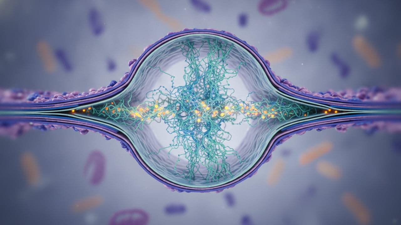
Researchers at Columbia University have developed three-dimensional stem cell models that recreate the initial structural formation of the human placenta. Described in a paper published in Nature Cell Biology, this biological invention allows scientists to observe how embryonic cells organize outside the womb. The platform offers a new path to examine why early implantation fails and how pregnancy complications emerge.
Suppressing Hippo signals to trigger placental tissue
Within days of fertilization, a human embryo makes its first developmental choice. Cells on the outer rim acquire an extraembryonic identity that instructs them to create the chorion, the early structural foundation of the placenta. Because implantation takes place inside the uterus and differs fundamentally from development in mice, investigating these early steps has posed a long-standing challenge.
To recreate the process in vitro, a research team led by Mijo Simunovic, assistant professor at Columbia University, focused on the Hippo signaling pathway. Known for regulating mechanical force sensing and cell growth, this biochemical pathway also directs the embryo's earliest specialization. According to reporting from MedicalXpress, researchers briefly reset human pluripotent stem cells to a naive state of potency, then administered chemical signals that suppressed Hippo activity to activate the placenta program.
“What is remarkable is that we don't have to tell each cell what to become or where to go.”
— Mijo Simunovic, Assistant Professor, Columbia University
Three weeks to form layered organoids
Once chemical cues initiated the placental sequence, the cells guided their own tissue assembly. Over three weeks, the 3D cell cultures produced the primary cell types found in the human chorion.
Without ongoing manual intervention, the cells organized into chorionic layers and generated fluid-filled spaces that mimic placental lacunae. In a developing pregnancy, these cavities appear during uterine implantation and later connect with maternal blood vessels. The autonomous patterning confirms that early extraembryonic cells retain the structural instructions needed to construct placental architecture.
Investigating pregnancy disorders and therapeutic avenues
Structural disruptions during early placenta development can lead to pregnancy loss and conditions like preeclampsia and fetal growth restriction, both of which cause maternal and neonatal deaths. While the study led by Miaoci Zhang does not list an associated patent application, the laboratory innovation gives scientists an experimental system tailored specifically to human tissue.
Simunovic and his colleagues will use the 3D organoid system to evaluate how physical forces and biochemical signals govern placenta assembly. Future experiments will also model interactions between fetal structures and maternal tissue to support fertility diagnostics and clinical treatments.
Read also on The Patent: Why ASPM Mutations Strike the Human Brain Harder Than Other Mammals · Can robots feel empathy? According to Columbia University yes